Diagnosis, diabetes, and vitreoretinal surgery

Retina & Vitreous

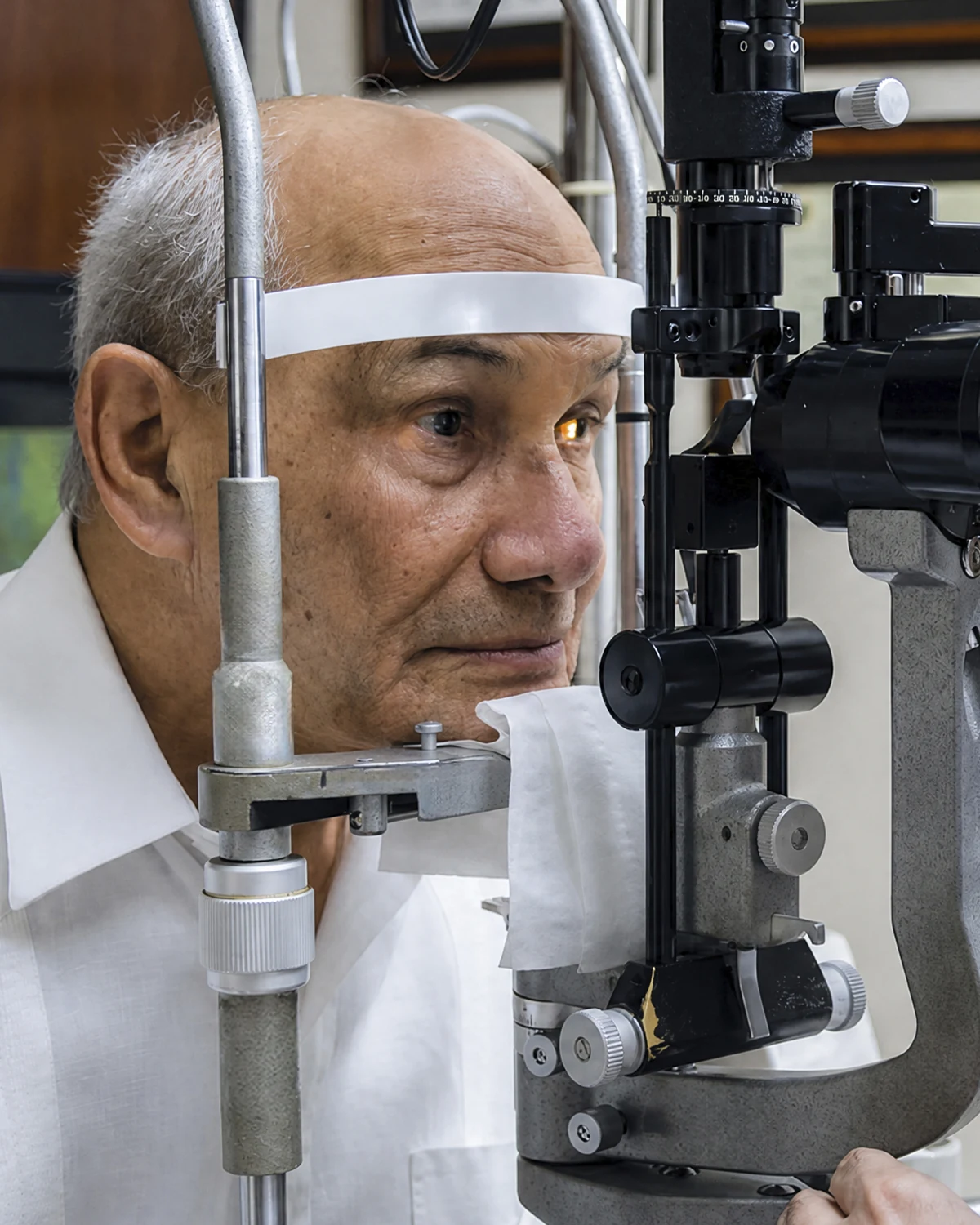
The retina is where everything you see comes into focus. If you have diabetes or high myopia, or you suddenly notice flashes and floaters, you need to be seen right away. We treat diabetic retinopathy, macular degeneration, and retinal detachment with precise diagnosis and vitreoretinal surgery.

Retina diagnosis and surgery at Centro Láser, Santo Domingo

What you should know about the retina

The retina is the layer of nerve tissue lining the back of the eye. Think of it as camera film: it captures light and converts it into the signals your brain reads as images. The macula, its central zone, is responsible for sharp, detailed vision. When the retina is affected by disease, vision loss can be severe and, in many cases, permanent if it isn't treated in time.

The most common retinal diseases are tied to broader health conditions. Diabetic retinopathy is the leading cause of preventable blindness in working-age adults, and age-related macular degeneration is the top cause of central vision loss after age 60. Retinal detachment, vascular occlusions, epiretinal membrane, and macular hole are also common.

Many of these diseases progress silently in their early stages. That's why, if you have diabetes, an annual dilated eye exam isn't optional: it's the tool that catches and treats retinopathy before it damages your vision. And if you notice flashes of light, a sudden shower of floaters, or a shadow across your field of vision, you need care right away.

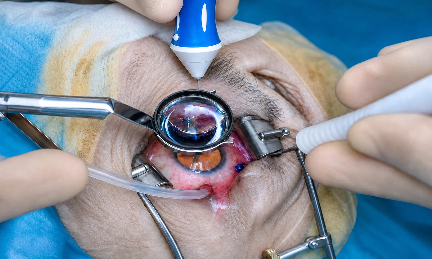
At Centro Láser, our retina and vitreous department combines precision diagnostics (OCT, angiography, and retinal photography) with the full range of treatments: intravitreal anti-VEGF injections, laser photocoagulation, vitrectomy, and retinal detachment surgery, backed by emergency care for the cases that can't wait.
What we
address

Each condition has its own symptoms, diagnosis, and treatment. If you recognize your symptoms in one of them, we can help.

Diabetic Retinopathy

The damage diabetes causes to the blood vessels in the retina. It's the leading cause of preventable blindness in working-age adults. It progresses silently in its early stages, which is why every diabetic patient needs a dilated eye exam at least once a year, even with good vision.

  • You have diabetes (type 1 or type 2)
  • Blurry or fluctuating vision
  • Dark spots or floaters in your vision
  • Difficulty seeing at night
  • Blank spots or shadows in your field of vision

Macular Degeneration (AMD)

The deterioration of the macula, the central part of the retina responsible for sharp, detailed vision. It's the leading cause of central vision loss after age 60. The wet form progresses quickly but responds very well to anti-VEGF injections when treated in time.

  • Straight lines look wavy or bent
  • A blurry or blank spot in the center of your vision
  • Colors look less vivid
  • You need more light to read
  • You are 60 or older

Retinal Detachment

The retina pulls away from the back wall of the eye, cutting off vision in the affected area. This is a medical emergency measured in hours, not days. Without timely surgery, vision loss can be permanent. If you notice the symptoms, seek care right away.

  • Sudden flashes of light
  • A sudden shower of floaters
  • A shadow, curtain, or veil covering part of your vision
  • Rapid vision loss in one eye
  • High myopia or a recent blow to the eye

Epiretinal Membrane and Macular Hole

Changes in the macula that distort central vision: a thin membrane that forms over the retina, or a small hole at its center. Both are diagnosed with OCT and, when they affect vision, treated with vitrectomy with good results.

  • Distorted or blurry central vision
  • Straight lines look wavy
  • Difficulty reading or recognizing faces
  • A spot-like sensation in the center of your vision
  • Double vision in one eye

This is how the world looks
with macular degeneration

Slide the bar to compare. Macular degeneration erases the center of your vision: whatever you look at directly darkens or distorts, while peripheral vision is preserved.

When treatment
makes sense

You don't have to wait. Treatment is considered as soon as one or more of these signs appear.

  • You have diabetes (every diabetic patient needs a dilated eye exam at least once a year)

  • You notice new flashes of light or floaters, or a sudden increase in them

  • You see a shadow, curtain, or veil covering part of your field of vision

  • Straight lines look wavy, or you notice a blurry spot in the center of your vision

  • You have high myopia or a family history of retinal problems

How we treat
this condition

From medical treatment of diabetes and macular disease to complex vitreoretinal surgery. The full range of care for every retinal condition.

Intravitreal Injections (Anti-VEGF) Saves the macula

Intravitreal Injections (Anti-VEGF)

Anti-VEGF medication injected inside the eye to slow wet macular degeneration and diabetic macular edema. The world's go-to treatment.

Duration
10-15 minutes
Anesthesia
Topical (anesthetic eye drops)
Recovery
1-2 days to resume activities
Explore the procedure
Retinal Detachment Surgery Urgent

Retinal Detachment Surgery

Emergency surgery to reattach a detached retina. Pneumatic retinopexy, scleral buckle, or vitrectomy depending on your case. Timing is everything.

Duration
60-120 minutes
Anesthesia
Local anesthesia with sedation or general anesthesia
Recovery
30 days
Explore the procedure
Retinal Laser Photocoagulation Prevents blindness

Retinal Laser Photocoagulation

Laser treatment that seals leaking vessels and reinforces retinal tears. Prevents diabetic retinopathy and retinal detachment from progressing.

Duration
15-30 minutes
Anesthesia
Topical (anesthetic eye drops)
Recovery
1-2 days to resume activities
Explore the procedure
Vitrectomy Vitreoretinal Surgery

Vitrectomy

Microscopic surgery that removes the vitreous gel to repair the retina. Treats vitreous hemorrhage, epiretinal membrane, macular hole, and advanced diabetic eye disease.

Duration
45-90 minutes per eye
Anesthesia
Local anesthesia with sedation, or general anesthesia
Recovery
21 days
Explore the procedure

Your recovery,
step by step.

Visual recovery is quick. We're with you at every post-op check until you're discharged.

01 /03

Injection or laser

Intravitreal injections and laser photocoagulation are outpatient procedures. You go home the same day with mild discomfort or temporary blurry vision that clears within hours.

Day of the procedure
02 /03

Vitrectomy recovery

After a vitrectomy or detachment surgery, if gas is used you may need to hold a specific head position for several days. Vision returns gradually.

Weeks (surgery)
03 /03

Ongoing monitoring

Many retinal diseases (diabetes, AMD) are chronic and require periodic checkups and, sometimes, maintenance injections to preserve your vision.

Follow-up

Why choose us
for your care

Four concrete reasons to trust the health of your eyes to this team.

24/7

Retina emergency care

Retinal detachment is an emergency measured in hours, not days. We run an emergency service ready to treat these cases when every moment counts.

OCT

Precision diagnostics

Optical coherence tomography (OCT), angiography, and retinal photography let us see the retina layer by layer and catch disease at its earliest stages.

Anti VEGF

Diabetes and macular treatment

Intravitreal anti-VEGF injections, the worldwide standard for wet macular degeneration and diabetic macular edema, given in the operating room under a sterile protocol.

Vitreous

Complete vitreoretinal surgery

Vitrectomy and retinal detachment surgery performed by fellowship-trained surgeons, for cases that require surgical intervention.

The technology behind
every procedure

Every pre-surgical study in one place, without ever leaving the center.

OCT Optical coherence tomography of the retina and optic nerve to catch silent diseases
Fluorescein Angiography Study of the retinal blood vessels with contrast dye
B-Scan Ultrasound Imaging of the eye's internal structures when the media are cloudy
Posterior Segment Photo Retinal photography: documents the condition of your retina

Specialized ophthalmologists
in the retina

Meet the team who will handle your evaluation, surgery, and follow-up.

How to book your visit

Four steps.
One visit.

We designed every step so you reach your treatment plan without unnecessary detours.

1 /04

Book your visit

By WhatsApp, form, or phone. Within 24 hours we confirm the day, time, and specialist at the location you prefer.

24 h
2 /04

Initial evaluation

A complete eye exam with your specialist. We talk through your history, symptoms, and what you want to achieve.

45 min
3 /04

Diagnostic testing

Whatever your case calls for, done inside the center and often during the same visit.

Same day
4 /04

Treatment plan

Your doctor walks you through options, costs, and next steps. You decide with clear information.

Next steps

We accept the country's
major health plans

Most plans cover surgery with standard options. Premium options may require a copay.

Learn more
about the retina

Patient guides reviewed by our specialists.

About
the retina

Yes, without exception. Diabetic retinopathy progresses without symptoms in its early stages: by the time your vision blurs, the damage can already be significant. That's why every diabetic patient needs a dilated eye exam at least once a year, even with perfect vision. It's the only way to catch and treat the damage in time. Diabetic retinopathy is the leading cause of preventable blindness in working-age adults, and preventing it depends on these checkups.

It can be. A sudden onset of flashes and a sharp increase in floaters can signal a retinal tear or detachment, which is a medical emergency. If you also notice a shadow or curtain covering part of your vision, seek care right away. Retinal detachment is measured in hours: treated in time, vision is preserved; delayed, the damage can be permanent.

They involve delivering a medication, usually an anti-VEGF drug, directly into the eye to treat wet macular degeneration, diabetic macular edema, and vascular occlusions. It sounds scarier than it is: the procedure is done in the operating room with numbing eye drops and a sterile protocol, takes just a few minutes, and most patients report little to no discomfort. It's the worldwide standard treatment for these diseases and has saved the vision of millions of people.

It depends on your disease and how your retina responds. Many conditions (wet AMD, diabetic edema) are chronic and require an initial loading schedule followed by maintenance doses spaced according to your response. Your retina specialist adjusts the frequency based on your OCT at each visit. This isn't a one-time treatment; it's ongoing management to preserve your vision long term.

It's the surgery that removes the vitreous (the clear gel that fills the eye) to access and repair the retina. It's used for retinal detachment, vitreous hemorrhage, epiretinal membrane, and macular hole. It's performed with microscopic instruments, and depending on the case, gas or silicone oil is placed inside the eye to hold the retina in position while it heals. It's a delicate surgery that requires a fellowship-trained vitreoretinal surgeon.

When a gas bubble is used to hold the retina in position, your surgeon may ask you to keep a specific head position for several days (for example, face down). The gas reabsorbs on its own over time. While you have the bubble, you cannot fly or travel to high altitudes, since the pressure change can be dangerous. Your care team will give you specific instructions for your case.

The dry form of AMD has no cure, but its progression can be slowed with lifestyle changes, risk factor control, and, in some cases, specific supplements. The wet form, which is more aggressive, responds very well to anti-VEGF injections: they stop the bleeding and, when treated in time, preserve or even improve vision in many patients. Early detection with OCT is key for both forms.

No. Most floaters are harmless and come from the natural aging of the vitreous. The challenge is telling the harmless ones apart from those that signal a retinal tear. The practical rule: if your floaters appear suddenly, increase a lot, or come with flashes of light or a shadow, you need an urgent evaluation. If they are few, stable, and long-standing, they are usually harmless, but it's worth confirming at a visit.

Optical coherence tomography (OCT) is a non-invasive test that images the retina layer by layer, with microscopic detail, in seconds and without touching the eye. It is the central tool of modern retina care: it detects macular edema, membranes, holes, and degeneration at very early stages, and lets your doctor precisely measure how you're responding to treatment at each visit. That's why it guides decisions about injections and surgery.

Where to
find us

Centro Láser Naco

C/ Fantino Falco #3, Ensanche Naco, Santo Domingo
+1 809 563-1324
Mon-Fri 7:30am – 5:00pm | Sat 7:30am – 12:00pm

Centro Láser Megacentro

Av. San Vicente de Paúl 321, Megacentro, Santo Domingo Este
+1 809 563-1324
Mon-Fri 7:30am – 5:00pm | Sat 7:30am – 12:00pm

Legacy Eye Clinic

C. Ing. Juan Paradas Bonilla 11, Santo Domingo

Your retina can't wait: get it evaluated in time

An evaluation with a fellowship-trained retina specialist. Dilated eye exam, OCT, and complete testing to catch and treat what your vision isn't showing yet.