HomeProceduresVitrectomy

Retina & Vitreous

Vitrectomy

Surgery that removes the vitreous gel to access and repair the retina. Treats vitreous hemorrhage, epiretinal membrane, macular hole, and complex cases of advanced diabetic retinopathy.

Vitrectomy

What Is a Vitrectomy?

A vitrectomy is the surgery that removes the vitreous, the clear gel that fills the inside of the eye, so the surgeon can access and repair the retina. It’s the core procedure of the vitreoretinal subspecialty, and it treats conditions that don’t respond to medical management or that require direct intervention on the retina.

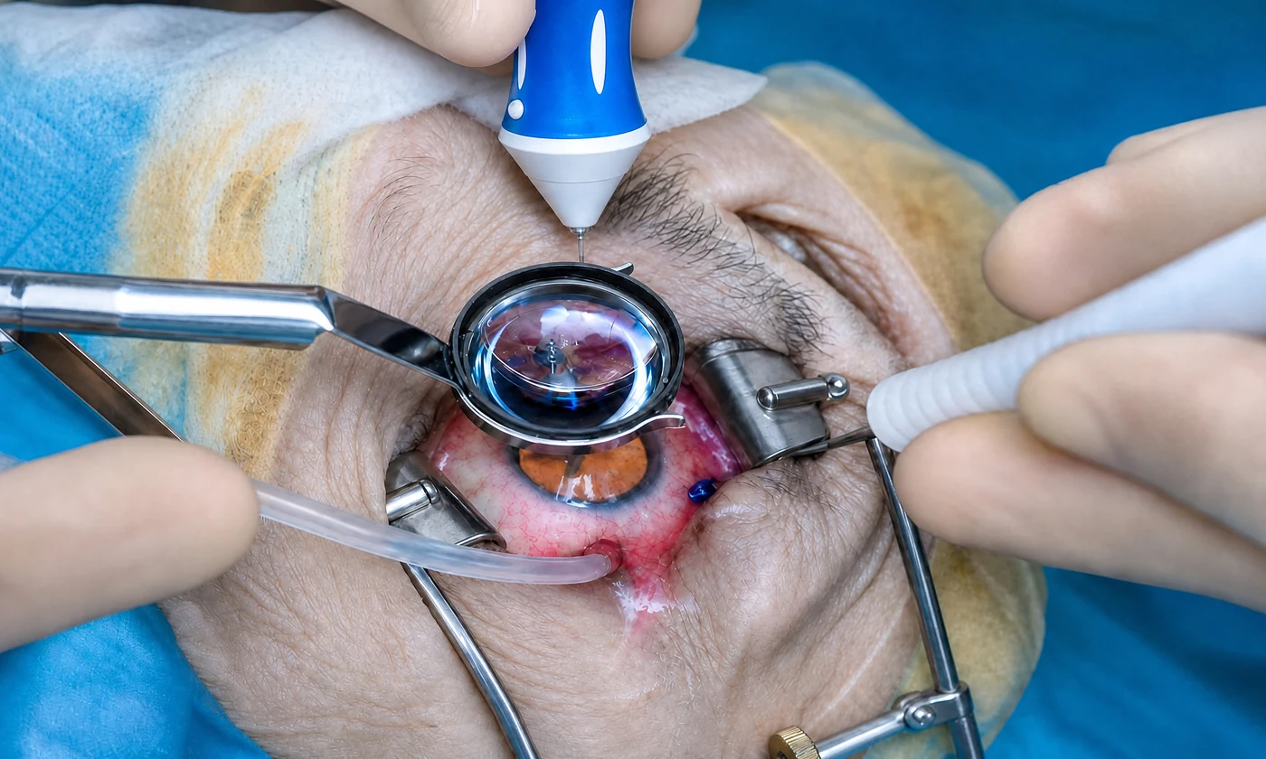
It’s performed with microscopic instruments inserted through tiny incisions in the pars plana, a safe zone of the eye. Once the vitreous is removed, the surgeon has direct access to the retina to peel membranes, close holes, drain blood, apply laser, or reattach a detached retina.

What Conditions Is It Used For?

  • Vitreous hemorrhage: blood inside the eye that doesn’t clear on its own, common in advanced diabetic retinopathy
  • Epiretinal membrane: a layer of tissue that forms over the macula and distorts vision
  • Macular hole: an opening in the center of the retina that affects fine, detailed vision
  • Advanced diabetic retinopathy with traction on the retina
  • Retinal detachment: to reattach the retina (see also retinal detachment surgery)

Gas and Silicone Oil

In many cases, once surgery is complete, the vitreous space is filled with a material that holds the retina in place while it heals:

  • Gas: absorbs on its own over time. Requires keeping your head in a specific position for several days and avoiding air travel until it’s gone.
  • Silicone oil: used for complex cases. Removed in a second, scheduled surgery.

Step by Step

  1. Full evaluation: OCT, ultrasound if the vitreous is cloudy, and surgical planning.
  2. Anesthesia: local with sedation, or general.
  3. Microscopic incisions in the pars plana and placement of the instruments.
  4. Vitreous removal and the specific repair needed (membrane peeling, hole closure, blood drainage, laser, retinal reattachment).
  5. Gas or oil fill if the case requires it.
  6. Closure and bandaging. The procedure takes 45 to 90 minutes.

Recovery

Visual recovery is gradual:

  • First few weeks: blurry vision, especially if gas was used (you’ll see the bubble move and shrink)
  • Discomfort, irritation, and a gritty feeling that ease with eye drops
  • Frequent follow-up visits to monitor healing

Full recovery can take anywhere from several weeks to a few months. It’s very common for a cataract to develop or progress after a vitrectomy, which is then treated later with standard cataract surgery.

Why Choose Centro Láser for Your Vitrectomy

Centro Láser has a retina department with fellowship-trained vitreoretinal surgeons and the equipment for modern microsurgery. We perform vitrectomy for the full range of indications, from epiretinal membrane to complex cases of retinal detachment and advanced diabetic retinopathy, with the long-term follow-up these surgeries require.


What you should know
about Vitrectomy

A vitrectomy removes the vitreous, the clear gel that fills the inside of the eye, so the surgeon can access and repair the retina. It's performed with microscopic instruments through tiny incisions. Once the vitreous is removed, the surgeon can peel membranes, close holes, apply laser, or drain blood, depending on the case. It's the core procedure of the vitreoretinal subspecialty.

For vitreous hemorrhage (bleeding inside the eye, common in diabetics), epiretinal membrane (a layer of tissue that distorts the macula), macular hole, advanced cases of diabetic retinopathy with traction, and retinal detachment. It's also used to remove the vitreous when there's an intraocular infection or other complications.

In many cases, yes. At the end of surgery, the vitreous space is filled with a bubble of gas (which absorbs on its own over time) or silicone oil (which is removed in a second surgery), to hold the retina in place while it heals. If you receive gas, you'll need to keep your head in a specific position for a few days and won't be able to fly until the bubble is gone.

Not during surgery. It's performed with local anesthesia and sedation or, in some cases, general anesthesia, so it's painless. Recovery tends to be more uncomfortable than painful: a gritty feeling, irritation, and blurry vision, all managed with eye drops and improving gradually.

Visual recovery is gradual and depends on the condition being treated and the retina's condition beforehand. In the first few weeks, vision is blurry, especially if gas was used (you'll see the bubble move). Vision improves as the gas absorbs and the retina heals. Full recovery can take anywhere from several weeks to a few months, with frequent follow-up visits.

That depends on how much damage the retina had before surgery. A vitrectomy repairs the structural problem (blood, membrane, hole, detachment), but the final visual outcome depends on the condition of the macula and optic nerve. The sooner the surgery is done, the better the prognosis tends to be. Your surgeon will walk you through realistic expectations for your specific case.

Like any intraocular surgery, it carries risks: cataract development or progression (common and treatable), increased eye pressure, infection (uncommon with sterile protocol), bleeding, or a new detachment. A fellowship-trained surgeon and close follow-up minimize these risks. The benefits of repairing the retina generally outweigh the risks by a wide margin in the cases where surgery is indicated.

Our specialists
in Vitrectomy

Are you a candidate for Vitrectomy?

Book your evaluation with our specialists.