When vision begins in the brain

Neuro-Ophthalmology

Not every vision problem starts in the eye. Some come from the optic nerve or the brain, and often the eye looks completely normal on exam. Neuro-ophthalmology connects your vision to your nervous system to find the cause when no one else can.

Neuro-ophthalmology and optic nerve care at Centro Láser, Santo Domingo

What you should know about neuro-ophthalmology

Neuro-ophthalmology is the subspecialty that studies how the eye and the brain work together. Vision doesn't end at the eye: visual information travels along the optic nerve, through the optic chiasm, and along the visual pathways all the way to the brain's visual cortex. A problem anywhere along that path can affect your vision, even when the eye itself is perfectly healthy.

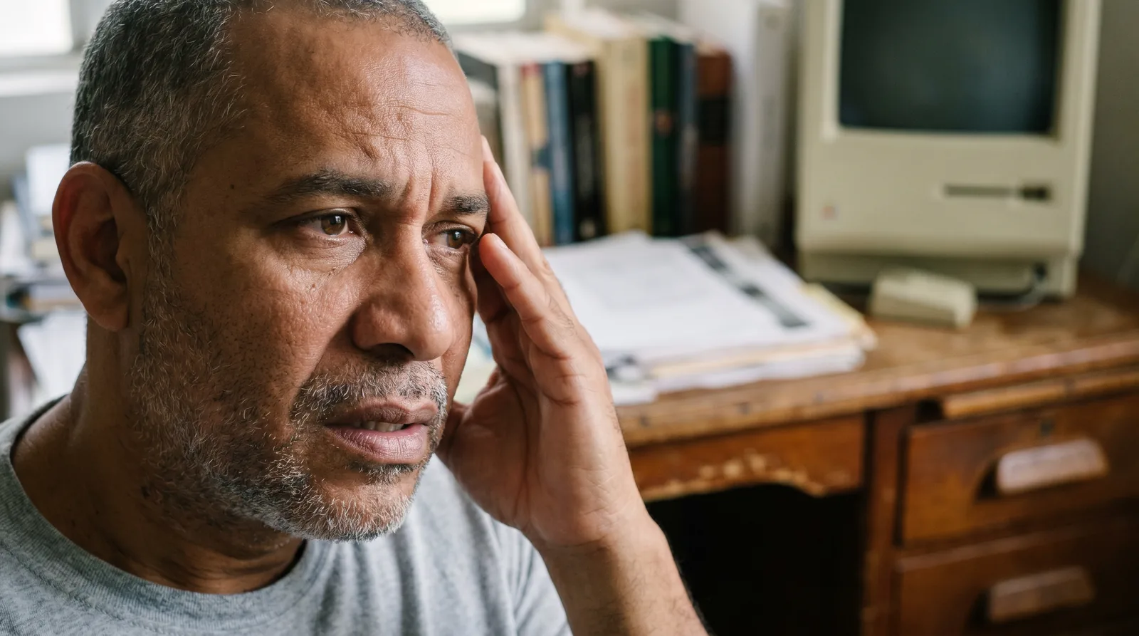
That explains a frustrating, sometimes frightening situation: someone loses vision, starts seeing double, or notices something off in their visual field, sees an eye doctor, and hears, "your eye looks fine." And that's often true. The problem isn't in the eye; it's in the optic nerve or the brain. That's where a neuro-ophthalmologist comes in, working like a detective for vision.

We treat conditions like paralysis of the nerves that move the eye (which cause double vision), optic nerve disease, chiasm compression from pituitary tumors, visual field changes caused by strokes, and pupil disorders like Horner syndrome. Many of these conditions are the first sign of a neurological disease that needs attention.

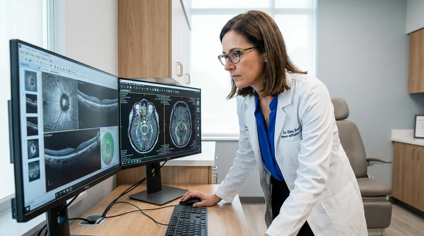
Our approach combines a thorough neuro-ophthalmic evaluation, testing like computerized visual field exams and optic nerve OCT, neuroimaging coordination when it's needed, and diplopia management for double vision and eye muscle paralysis, always working alongside neurology and other specialists.
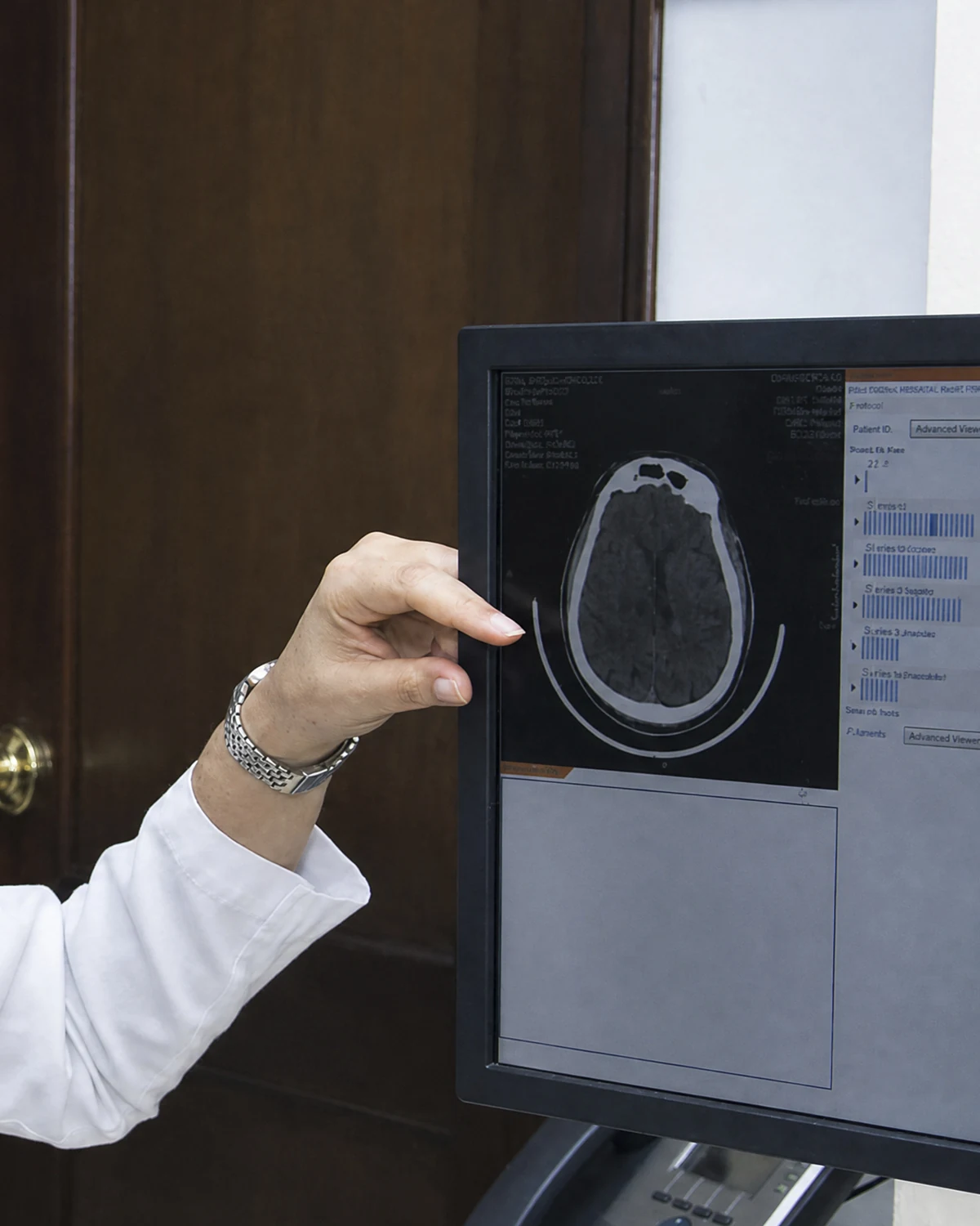
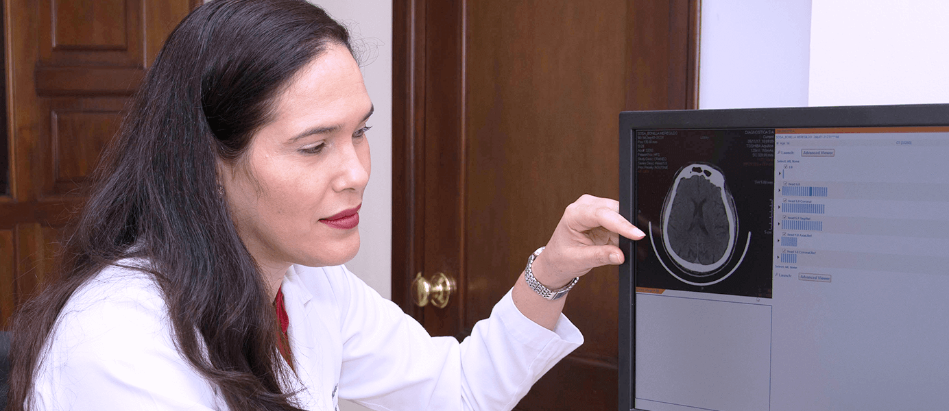
What we
address

Each condition has its own symptoms, diagnosis, and treatment. If you recognize your symptoms in one of them, we can help.

Double Vision (Diplopia) and Eye Muscle Palsy

When the nerves that move your eyes are affected, your eyes lose coordination and you start seeing double. This can result from eye muscle palsy, trauma, aneurysms, or multiple sclerosis. New-onset double vision should always be evaluated, since it can sometimes signal a neurological problem that needs attention.

  • You see two images of the same object
  • The double vision goes away when you cover one eye
  • You tilt or turn your head to see better
  • A recently drooping eyelid
  • Sudden onset, sometimes with a headache

Optic Nerve Disease

The optic nerve carries information from the eye to the brain. When it becomes inflamed, loses blood supply, or is affected by a neurological disease, vision drops even though the rest of the eye is healthy. Early diagnosis with visual field testing and OCT is key to protecting the vision you have left.

  • Vision loss in one eye, sudden or gradual
  • Colors look dull or washed out
  • Pain when moving the eye (in some cases)
  • A blurry spot or dark area in your vision
  • A known neurological condition

Visual Field Defects and Chiasm Compression

Pituitary tumors can compress the optic chiasm, and strokes can damage the visual pathways, causing loss of specific parts of your visual field, like half of your vision on one side. Patients often don't notice until they start bumping into things. Visual field testing picks it up.

  • You bump into objects or people on one side
  • A sense that part of your vision is missing
  • Loss of side vision in both eyes
  • Headaches that come with it
  • A history of a pituitary tumor or stroke

Pupil Disorders (Horner Syndrome)

Your pupils reflect the state of your nervous system. A difference in size between them, or a drooping eyelid alongside it, can point to Horner syndrome or another neurological issue. It's sometimes benign, but a neuro-ophthalmic evaluation determines whether there's an underlying cause that needs attention.

  • One pupil smaller or larger than the other
  • A slightly drooping eyelid on the same side
  • A difference that shows up recently
  • Uneven sweating on one side of the face
  • Sometimes it's first noticed in a photo or pointed out by someone else

This is how the world looks
with hemianopia

Slide the bar to compare. When the damage is in the brain or the visual pathways, half of the visual field can be lost, and many people do not notice until they bump into things on one side.

When treatment
makes sense

You don't have to wait. Treatment is considered as soon as one or more of these signs appear.

  • You've lost vision, suddenly or gradually, with no clear eye-related cause

  • You're seeing double (one object looks like two)

  • You notice part of your visual field is missing, or you keep bumping into things on one side

  • You have a recently drooping eyelid or one pupil that's larger than the other

  • You have headaches with visual symptoms, or you were referred by neurology

How we treat
this condition

Precision diagnosis and management for conditions where vision and the nervous system meet, in coordination with neurology.

Diplopia Management Single Vision

Diplopia Management

We diagnose the cause of double vision and treat it with prisms, occlusion, or management of the underlying condition, restoring single vision.

Duration
45-60 minute evaluation
Anesthesia
Not required
Recovery
1-2 days to resume activities
Explore the procedure
Neuro-ophthalmic Evaluation Precision Diagnosis

Neuro-ophthalmic Evaluation

Studies the entire visual pathway, from eye to brain, combining exam, visual field testing, OCT, and neuroimaging. Finds the cause a normal eye exam can't explain.

Duration
60-90 minutes
Anesthesia
Not required
Recovery
1-2 days to resume activities
Explore the procedure

Your recovery,
step by step.

Visual recovery is quick. We're with you at every post-op check until you're discharged.

01 /03

A thorough workup

The neuro-ophthalmic evaluation is thorough: it combines your history, the exam, visual field testing, and OCT to map out where the problem is along the visual pathway.

The evaluation
02 /03

Neuroimaging and neurology

Depending on what we find, we coordinate an MRI or other imaging and work alongside neurology to confirm the underlying cause.

Coordination
03 /03

Treatment and follow-up

Treatment depends on the cause, anything from managing diplopia with prism lenses to ongoing optic nerve monitoring, coordinated with the rest of your medical team.

Management

Why choose us
for your care

Four concrete reasons to trust the health of your eyes to this team.

Eye+

Vision beyond the eye

We evaluate the entire visual pathway, from the eye to the brain. When a normal eye exam doesn't explain your symptoms, this is where we look for the real cause.

Detect

Diagnosing the hard cases

Neuro-ophthalmology is precision diagnosis: piecing together symptoms, visual field testing, OCT, and neuroimaging to find the cause when others can't.

Network

Partnership with neurology

Many neuro-ophthalmic conditions are the first sign of a neurological disease. We work closely with neurology and other specialists to get you answers.

1

Testing under one roof

Computerized visual field testing, optic nerve OCT, and specialized evaluation, all at the same eye center, with neuroimaging coordinated for you.

The technology behind
every procedure

Every pre-surgical study in one place, without ever leaving the center.

OCT Optical coherence tomography of the retina and optic nerve to catch silent diseases
Humphrey Visual Field Measures your peripheral vision to detect glaucoma or neurological problems

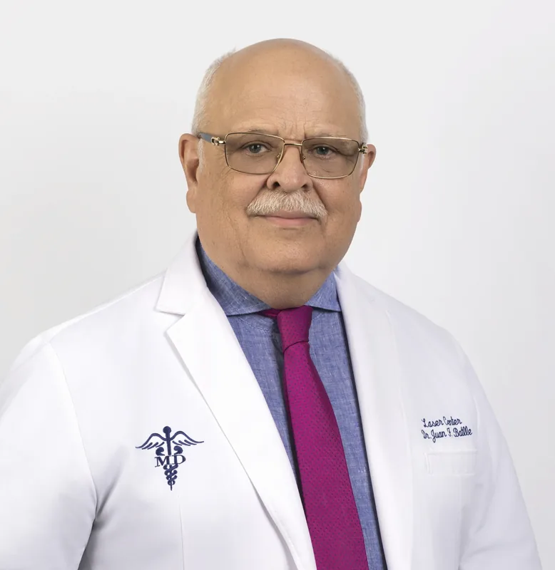
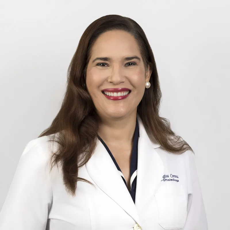
Specialized ophthalmologists
in neuro-ophthalmology

Meet the team who will handle your evaluation, surgery, and follow-up.

How to book your visit

Four steps.
One visit.

We designed every step so you reach your treatment plan without unnecessary detours.

1 /04

Book your visit

By WhatsApp, form, or phone. Within 24 hours we confirm the day, time, and specialist at the location you prefer.

24 h
2 /04

Initial evaluation

A complete eye exam with your specialist. We talk through your history, symptoms, and what you want to achieve.

45 min
3 /04

Diagnostic testing

Whatever your case calls for, done inside the center and often during the same visit.

Same day
4 /04

Treatment plan

Your doctor walks you through options, costs, and next steps. You decide with clear information.

Next steps

We accept the country's
major health plans

Most plans cover surgery with standard options. Premium options may require a copay.

Learn more
about neuro-ophthalmology

Patient guides reviewed by our specialists.

About
neuro-ophthalmology

It's the subspecialty that studies how the eye and the brain work together. Vision doesn't end at the eye: visual information travels along the optic nerve and the visual pathways all the way to the brain. Neuro-ophthalmology deals with vision problems that start along that path, not in the eye itself, things like optic nerve disease, double vision from eye muscle palsy, or visual field defects caused by neurological conditions.

It's more common than it sounds, and it's exactly why neuro-ophthalmology exists. If the problem is in the optic nerve or the brain, the eye can look perfectly normal on a routine exam and your vision can still be failing. A routine eye exam doesn't evaluate the entire visual pathway. A neuro-ophthalmologist studies that part of the path to find the cause a normal eye exam can't explain.

New-onset double vision (diplopia) should always be evaluated, especially if it goes away when you cover one eye, which points to a problem with eye alignment or the nerves that move the eye. It can have treatable causes, but it can also signal a neurological problem that needs attention, like a cranial nerve palsy. Don't wait it out: an evaluation will tell you the cause and how urgent it is.

The main ones are computerized visual field testing (which maps out which parts of your vision are working and which aren't), optic nerve OCT (which measures the health of the nerve fibers), and, when needed, an MRI of the brain and visual pathways. The key is putting these tests together with your history and exam to reach a diagnosis. Neuro-ophthalmology is, above all, precision diagnosis.

Because when we suspect the cause of a vision problem lies in the brain, the visual pathways, or structures like the pituitary gland, an MRI lets us see them in detail. For example, a pituitary tumor can compress the optic chiasm and cause visual field loss. An MRI confirms or rules out these causes. We coordinate it and read it together with neurology when needed.

It's the loss of part of what you see while other parts stay intact. For example, losing half of your visual field, on one side, in both eyes. Patients often don't notice it directly; instead, they bump into objects or people on one side. These defects follow characteristic patterns depending on where the problem sits along the visual pathway, so visual field testing doesn't just detect it; it helps pinpoint the cause.

Not always, but it's worth getting checked. A difference in pupil size (anisocoria) can be a normal variant in some people, but it can also point to Horner syndrome or another neurological issue, especially if it's new or comes with a drooping eyelid. A neuro-ophthalmic evaluation determines whether it's benign or whether there's a cause that needs attention.

Yes, closely. Many neuro-ophthalmic conditions are the first sign of a neurological disease (multiple sclerosis, tumors, aneurysms, stroke). The neuro-ophthalmologist is often the one who catches the visual clue and works together with neurology and other specialists to confirm the diagnosis and coordinate treatment. It's teamwork between the eye and the nervous system.

Anyone with vision loss that has no clear eye-related cause, double vision, visual field defects, a new drooping eyelid or unequal pupils, or visual symptoms that come with headaches. Also anyone referred by neurology to have the optic nerve or visual field evaluated. If a normal eye exam doesn't explain your symptoms, it's time to look beyond the eye.

Where to
find us

Centro Láser Naco

C/ Fantino Falco #3, Ensanche Naco, Santo Domingo
+1 809 563-1324
Mon-Fri 7:30am – 5:00pm | Sat 7:30am – 12:00pm

Centro Láser Megacentro

Av. San Vicente de Paúl 321, Megacentro, Santo Domingo Este
+1 809 563-1324
Mon-Fri 7:30am – 5:00pm | Sat 7:30am – 12:00pm

Legacy Eye Clinic

C. Ing. Juan Paradas Bonilla 11, Santo Domingo

When vision fails but the eye is healthy

Get evaluated by our neuro-ophthalmology team. We study the entire visual pathway, from the eye to the brain, to find the cause and coordinate your care.