Diagnosis, keratoconus, and transplant

Cornea

The cornea is the eye's most powerful lens, and a lot of vision loss starts there before you ever notice it. We treat keratoconus at every stage, along with corneal dystrophies and scarring, with a full range of options from cross-linking to corneal transplant.

Cornea and keratoconus treatment at Centro Láser, Santo Domingo

What you should know about the cornea

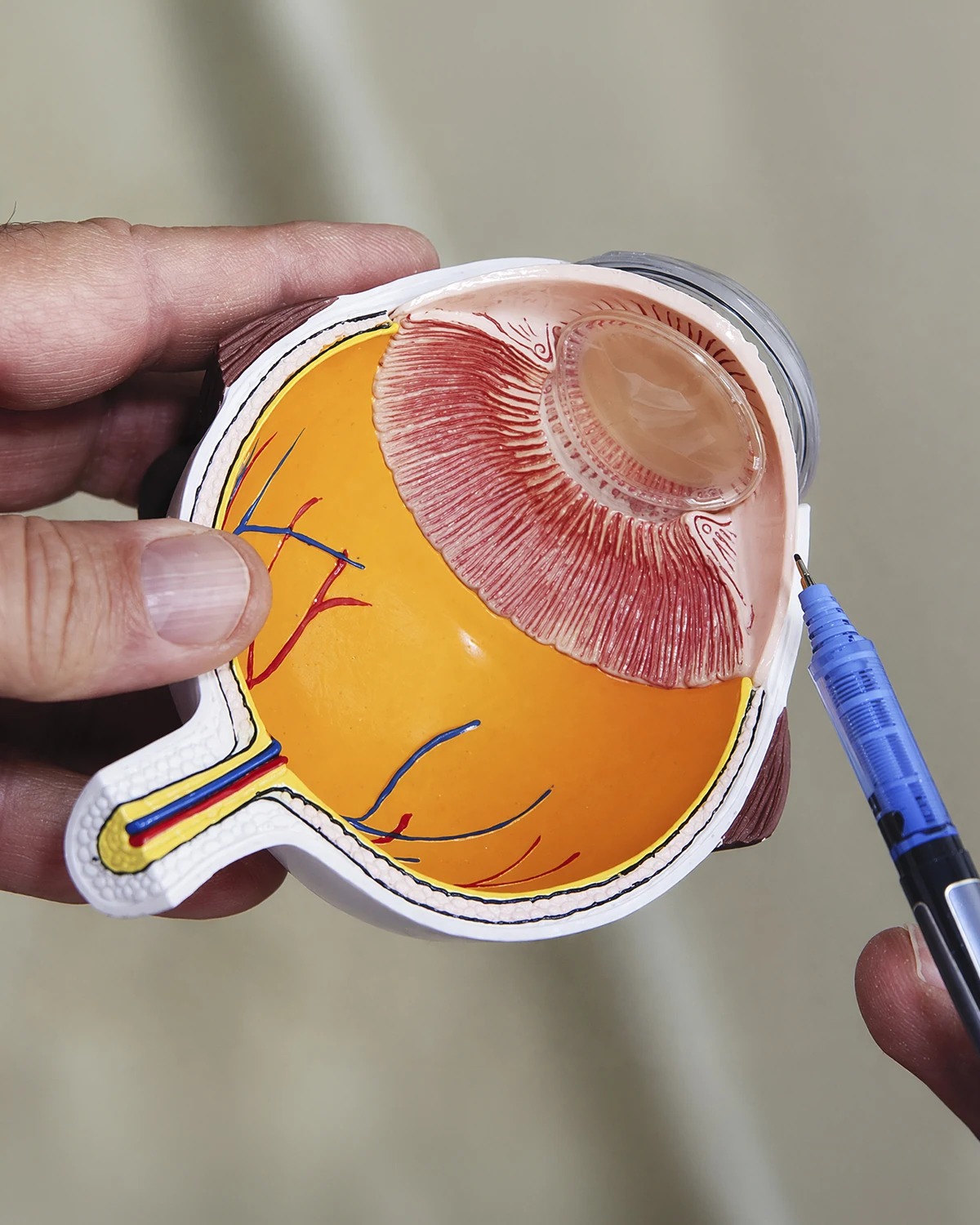
The cornea is the clear, curved surface at the front of the eye. It works as the most powerful lens in your visual system, focusing light onto the retina and protecting the eye's inner structures. When it loses its shape, clarity, or integrity, vision becomes distorted, and without treatment the damage can become permanent.

The most common corneal condition is keratoconus, a progressive disease that thins and reshapes the cornea, usually starting in the teenage years. Other conditions include inherited dystrophies, scarring or infection-related opacities, degenerations, and ectasia that can develop after refractive surgery.

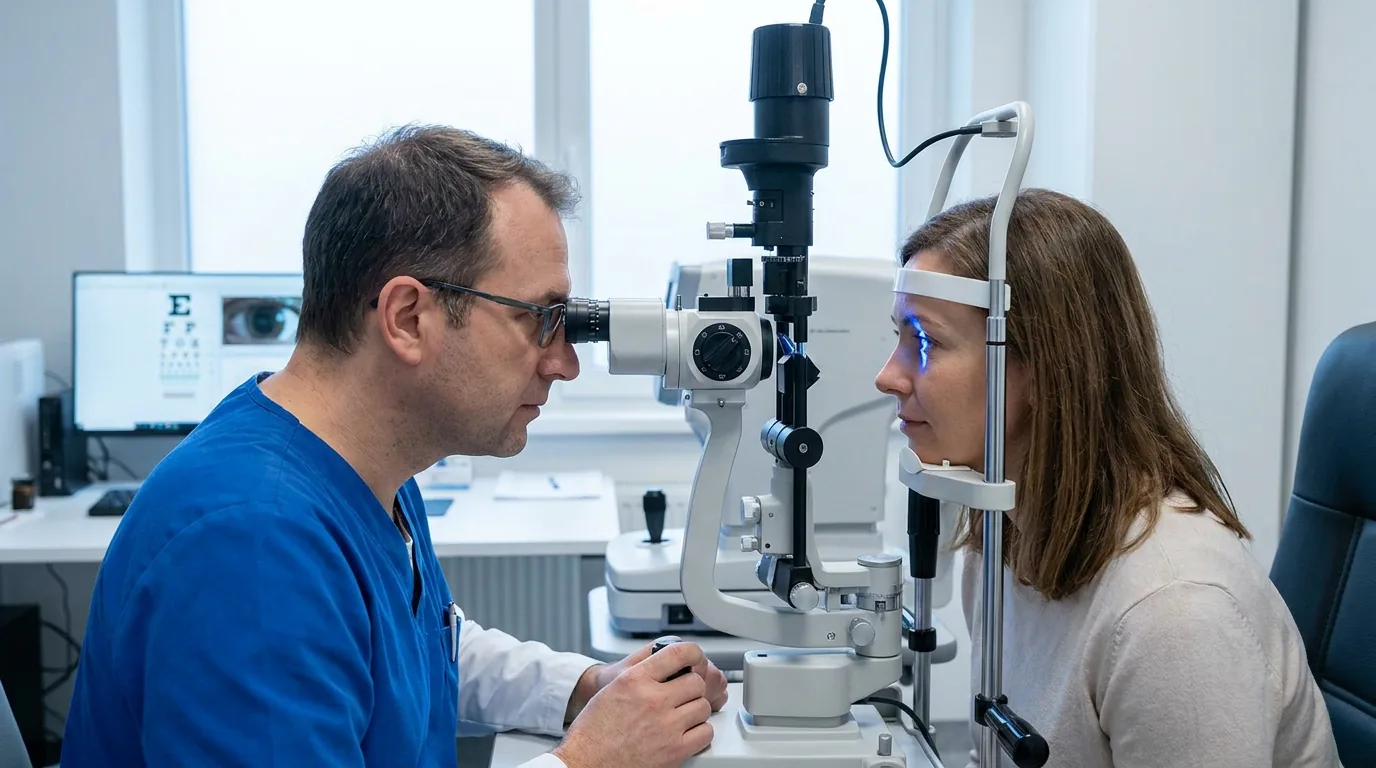
Our cornea department includes four fellowship-trained specialists and precision diagnostics: Pentacam corneal topography, specular microscopy, anterior segment OCT, and pachymetry. This lets us catch keratoconus at its earliest stages, while it can still be stopped in its tracks.

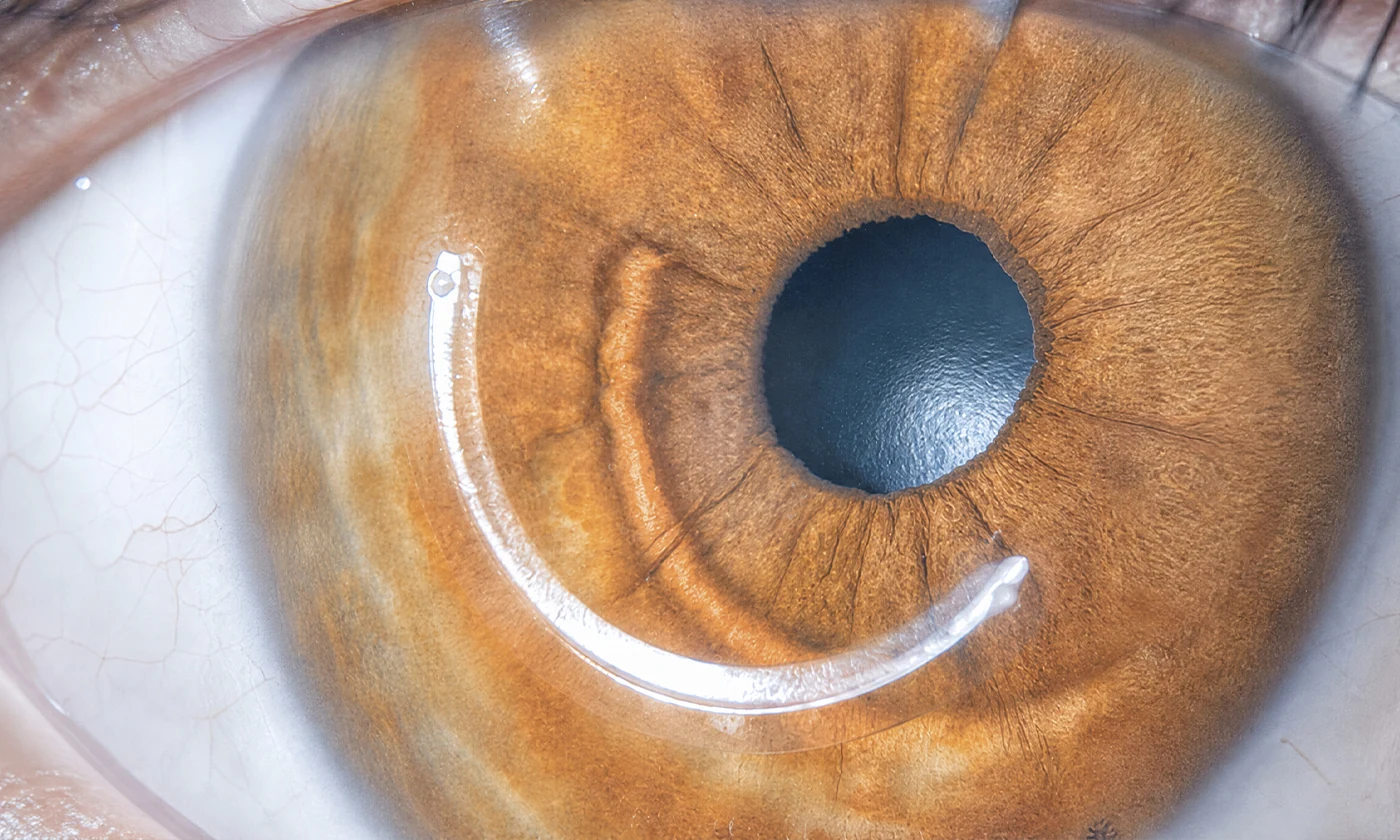
Treatment follows a step-by-step approach based on the stage of disease: corneal cross-linking to halt progression, intracorneal ring segments to reshape the curvature, and, when the cornea is already severely damaged, corneal transplant using a range of techniques. For the most complex cases where a transplant is not an option, we offer K-PRO keratoprosthesis.
What we
address

Each condition has its own symptoms, diagnosis, and treatment. If you recognize your symptoms in one of them, we can help.

Keratoconus

The most common corneal condition. The cornea gradually thins and bulges into a cone shape. It usually starts in the teenage years and can progress into your 30s. Early detection lets us stop it before it causes permanent vision loss.

  • Distorted or "stretched" vision at any distance
  • Frequent, fast changes in your prescription, especially astigmatism
  • Light sensitivity and halos around lights
  • Vision that never fully clears up, even with glasses
  • Rubbing your eyes often

Corneal Dystrophies

A group of inherited conditions that cause abnormal deposits or changes in the layers of the cornea. They typically affect both eyes symmetrically and progress slowly. Some require an endothelial transplant once they compromise clarity.

  • Blurry vision that gradually worsens
  • Vision that is worse when you wake up and improves through the day
  • Light sensitivity and glare
  • Family history of corneal problems
  • Episodes of eye pain from recurrent erosions

Corneal Opacities and Scarring

Loss of corneal clarity from scarring caused by past infections, trauma, or surgery. Light cannot pass through properly, so vision clouds over. Depending on how deep the scarring goes, treatment ranges from lamellar or penetrating transplant to keratoprosthesis.

  • A visible whitish spot on the cornea
  • Cloudy vision in the affected eye
  • History of eye infection, ulcer, or trauma
  • Intense glare in bright light
  • Vision that does not improve with glasses or contact lenses

Keratitis and Infectious Ulcers

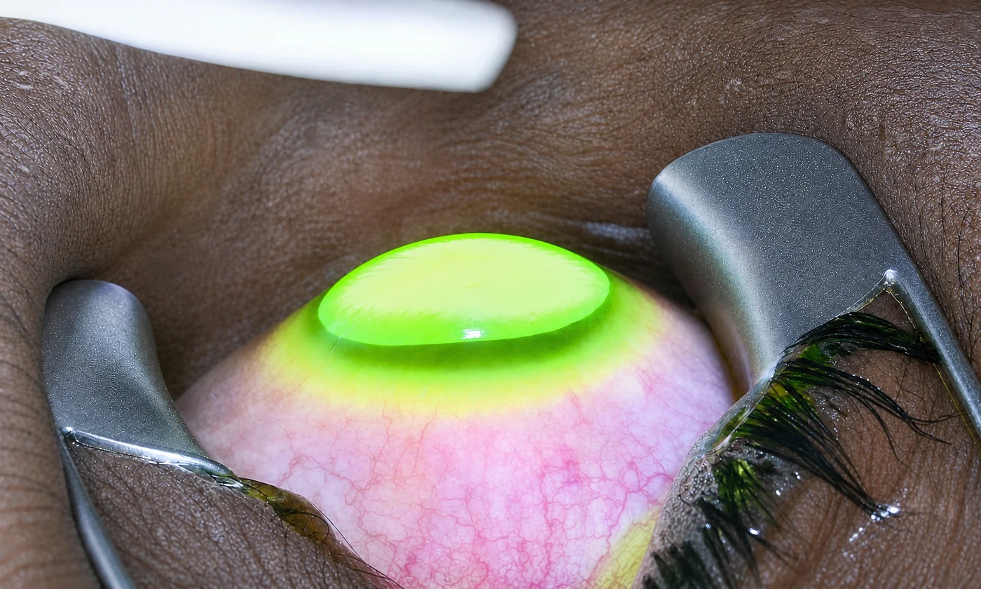
An infection of the cornea caused by bacteria, fungi, viruses, or parasites, common among contact lens wearers. It is an emergency: left untreated, it can leave permanent scarring or perforate the cornea. It needs immediate care.

  • Intense eye pain that comes on quickly
  • Marked redness and tearing
  • Extreme light sensitivity
  • Discharge and sudden blurry vision
  • Contact lens use, especially sleeping in them

This is how the world looks
with keratoconus

Slide the bar to compare. Keratoconus warps the cornea, producing blurry, distorted, ghosted vision that regular glasses can no longer fully correct.

When treatment
makes sense

You don't have to wait. Treatment is considered as soon as one or more of these signs appear.

  • You were diagnosed with keratoconus, or your astigmatism prescription keeps changing fast

  • Your vision is distorted or "stretched" and glasses do not fully correct it

  • You have a family history of keratoconus or corneal disease

  • You rub your eyes often and notice your vision getting worse

  • You have a corneal opacity, scar, or infection that is affecting your vision

How we treat
this condition

A complete treatment ladder based on the stage of your cornea, from stopping progression to restoring clarity.

Corneal Collagen Cross-Linking (CXL) Stops Keratoconus

Corneal Collagen Cross-Linking (CXL)

Stops keratoconus progression by strengthening the corneal collagen with riboflavin and UV light. The first line of defense to catch the disease early.

Duration
60-90 minutes
Anesthesia
Topical (anesthetic drops)
Recovery
7 days
Explore the procedure
Corneal Transplant (Keratoplasty) For advanced cases

Corneal Transplant (Keratoplasty)

Replaces a damaged or clouded cornea with healthy donor tissue. Modern techniques replace only the affected layer for a faster recovery.

Duration
45-90 minutes per eye
Anesthesia
Local anesthesia with sedation, or general anesthesia
Recovery
30 days
Explore the procedure
Intracorneal Ring Segments (ICRS) Reversible

Intracorneal Ring Segments (ICRS)

Segments that reshape the cornea's curvature in keratoconus. Placed with a femtosecond laser, they're reversible and improve vision without removing tissue.

Duration
15-20 minutes per eye
Anesthesia
Topical (anesthetic eye drops)
Recovery
7 days
Explore the procedure
Keratoprosthesis (Boston K-PRO) Complex cases

Keratoprosthesis (Boston K-PRO)

An artificial cornea for cases where a conventional transplant isn't viable or has failed. Restores vision in situations once considered untreatable.

Duration
60-120 minutes per eye
Anesthesia
Local plus sedation, or general anesthesia
Recovery
30 days
Explore the procedure

Your recovery,
step by step.

Visual recovery is quick. We're with you at every post-op check until you're discharged.

01 /03

After your corneal procedure

After cross-linking or ring placement, you will wear a bandage contact lens for the first few days, with light sensitivity and tearing that ease up with drops.

Days 1-7
02 /03

Visual recovery

Your vision stabilizes gradually. With rings, 80-90% of the improvement happens between weeks 2 and 4, with close follow-up visits.

Weeks 2-6
03 /03

Transplant follow-up

If you had a transplant, full visual recovery takes months and calls for long-term follow-up to watch for rejection and fine-tune your prescription.

Months (transplant)

Why choose us
for your care

Four concrete reasons to trust the health of your eyes to this team.

4

Specialists dedicated to cornea

An internationally trained cornea team, including transplant surgeons experienced in every modern technique.

Femto ®

Femtosecond laser ring placement

Intracorneal rings placed with femtosecond, micron-level precision, not by hand. Greater safety and consistency.

K-PRO

Keratoprosthesis for complex cases

We offer the K-PRO artificial cornea for patients where a conventional transplant isn't an option. Few centers in the region perform it.

1

A complete treatment ladder

From early cross-linking to advanced transplant surgery, all under one roof. We don't refer you elsewhere as your case progresses.

The technology behind
every procedure

Every pre-surgical study in one place, without ever leaving the center.

Pentacam Advanced corneal tomography: a detailed map of the shape of your cornea (Oculus)
Visante OCT Detailed imaging of the front of the eye (Zeiss)
Specular Microscopy Count and morphology of the corneal endothelium (Tomey / Konan)
Pachymetry Measures the thickness of your cornea to micrometer precision
Anterior Segment Photo Documents the condition of the iris, cornea, and lens
Topolyzer High-resolution corneal topography with a Placido disc camera

Specialized ophthalmologists
in the cornea

Meet the team who will handle your evaluation, surgery, and follow-up.

How to book your visit

Four steps.
One visit.

We designed every step so you reach your treatment plan without unnecessary detours.

1 /04

Book your visit

By WhatsApp, form, or phone. Within 24 hours we confirm the day, time, and specialist at the location you prefer.

24 h
2 /04

Initial evaluation

A complete eye exam with your specialist. We talk through your history, symptoms, and what you want to achieve.

45 min
3 /04

Diagnostic testing

Whatever your case calls for, done inside the center and often during the same visit.

Same day
4 /04

Treatment plan

Your doctor walks you through options, costs, and next steps. You decide with clear information.

Next steps

We accept the country's
major health plans

Most plans cover surgery with standard options. Premium options may require a copay.

Learn more
about the cornea

Patient guides reviewed by our specialists.

About
the cornea

It is the most common corneal disease. The cornea, normally round, gradually thins and bulges into a cone shape. This distorts your vision and creates an irregular astigmatism that keeps changing. It usually starts in the teenage years and can progress into your 30s. The good news: with early detection, we can stop it before it causes permanent vision loss.

With Pentacam corneal topography, a detailed map of your cornea's shape and thickness that can catch keratoconus at its earliest stages, before it affects your vision. That's why it's worth getting checked if your astigmatism is changing fast, you rub your eyes a lot, or it runs in your family. We round out the diagnosis with pachymetry, specular microscopy, and anterior segment OCT.

Yes. Corneal cross-linking stops progression in about 95% of cases. It uses riboflavin (vitamin B2) and ultraviolet light to strengthen the collagen fibers in the cornea, halting the reshaping process. It works best the earlier it's done, which is why early detection matters so much. It won't restore vision you've already lost, but it keeps keratoconus from advancing further.

Small, semicircular segments of biocompatible material inserted into the cornea to reshape its curvature and improve vision in patients with keratoconus. They are placed with a femtosecond laser, are minimally invasive, and, unlike a transplant, are reversible and adjustable. They are frequently combined with cross-linking to stabilize and improve vision at the same time.

Not necessarily. A transplant is reserved for advanced cases where the cornea is already severely reshaped, thinned, or clouded, and other options (cross-linking, rings, specialty lenses) are no longer enough. Most patients whose keratoconus is caught early never need a transplant. When one is needed, today's techniques can replace just the damaged layer, with a faster recovery than a full transplant.

It depends on which layer of the cornea is damaged. A penetrating transplant (PKP) replaces the full thickness; anterior lamellar (DALK) replaces the outer layers while keeping the innermost one; and endothelial transplants (DMEK and DSAEK) replace only the innermost layer, with faster recovery and a lower risk of rejection. Your surgeon chooses the technique based on your specific diagnosis.

Rejection is possible, but it is less common than with other organ transplants because the cornea has no blood vessels of its own. Modern techniques that replace only the damaged layer (DMEK, DSAEK) have even lower rejection rates. It is managed with eye drops and follow-up visits, and when caught early, most rejection episodes reverse. That is why long-term follow-up is essential.

It is an artificial cornea implanted when a conventional corneal transplant isn't an option, or has already failed more than once. It restores vision in very complex cases once considered untreatable. Few centers in the region perform this procedure. It's a resource for specific situations that your cornea specialist evaluates case by case.

Yes, especially if you sleep in them, wear them longer than recommended, or do not clean them properly. The main risk is infectious keratitis, a corneal infection that is a medical emergency: left untreated, it can leave permanent scarring. If you wear contact lenses and develop sudden intense eye pain, redness, and light sensitivity, seek care right away.

Where to
find us

Centro Láser Naco

C/ Fantino Falco #3, Ensanche Naco, Santo Domingo
+1 809 563-1324
Mon-Fri 7:30am – 5:00pm | Sat 7:30am – 12:00pm

Centro Láser Megacentro

Av. San Vicente de Paúl 321, Megacentro, Santo Domingo Este
+1 809 563-1324
Mon-Fri 7:30am – 5:00pm | Sat 7:30am – 12:00pm

Legacy Eye Clinic

C. Ing. Juan Paradas Bonilla 11, Santo Domingo

Your cornea has a solution at every stage

Meet with a fellowship-trained cornea specialist. Topography and a complete workup to confirm your diagnosis and find the right treatment for your case.