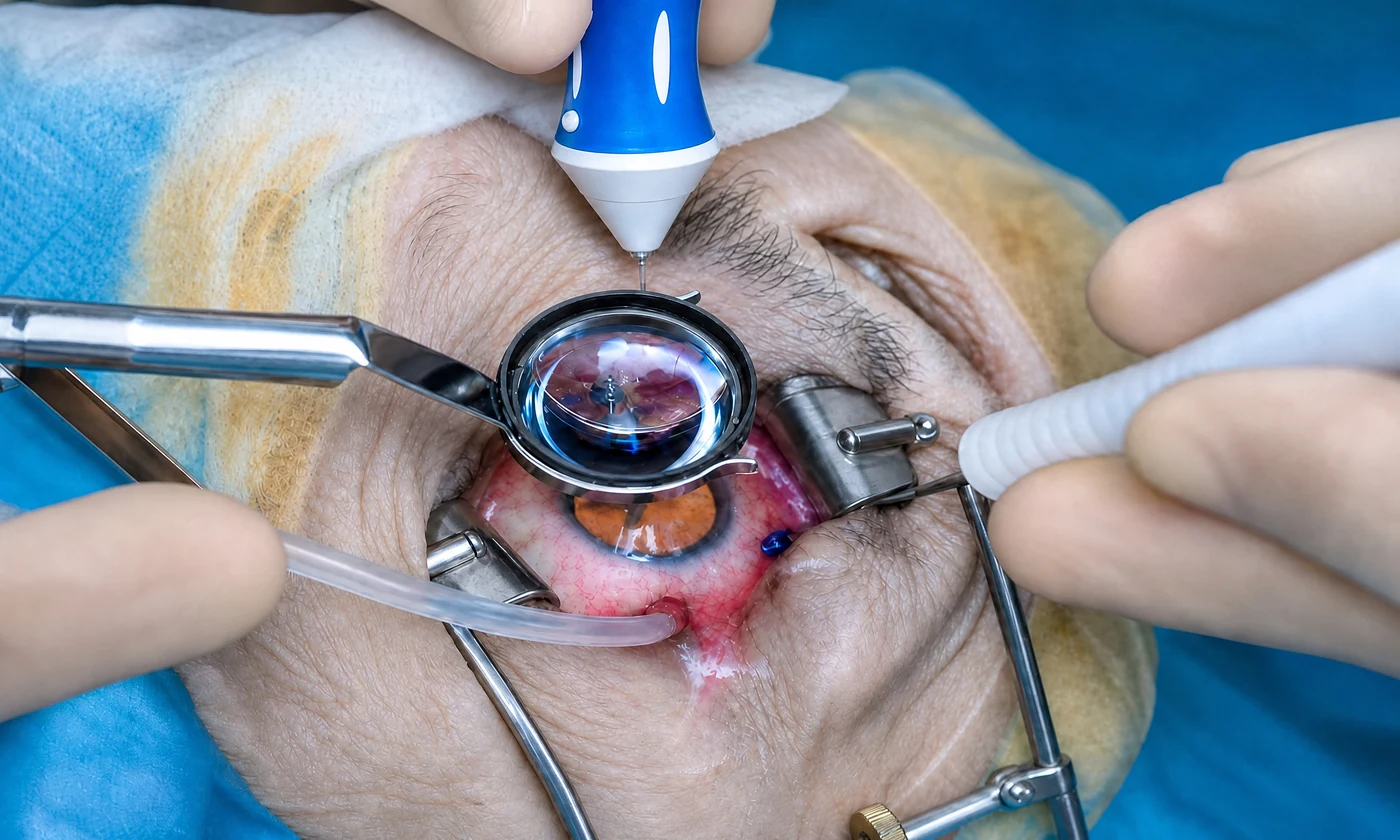
What Is Retinal Detachment Surgery?
Retinal detachment happens when the retina separates from the inner wall of the eye, cutting off vision in the affected area. It’s a medical emergency: once detached, the retina stops getting oxygen and its cells start to break down over time. The sooner it’s reattached, the better the visual outcome.
The goal of surgery is to reposition the retina and seal the tear or tears that caused the detachment. It’s one of the most time-sensitive surgeries in ophthalmology, so much so that outcomes are measured in hours.
Repair Techniques
The technique is chosen based on the type, location, and complexity of the detachment. They’re often combined.
Pneumatic Retinopexy
A gas bubble is injected into the eye to push the retina against the wall while it heals, combined with laser or cryotherapy to seal the tear. This is reserved for select cases, with tears in the upper part of the retina.
Scleral Buckle
A silicone band is placed around the outside of the eye to gently bring the eye wall closer to the retina, reducing traction and allowing it to reattach. It’s a well-established technique that works well for many types of detachment.
Vitrectomy
The vitreous gel is removed, the retina is reattached from the inside, sealed with laser, and the eye is refilled with gas or silicone oil to hold it in place. This is the preferred technique for complex detachments or those involving bleeding. See vitrectomy.
Why Timing Is Critical
The factor that most affects the visual outcome is whether the macula was detached and how much time has passed:
- Surgery before the macula detaches: the visual outlook is generally very good
- Surgery after the macula has detached: the retina can be reattached, but central vision may not fully recover
That’s why flashes of light, a sudden shower of floaters, or a shadow advancing across your field of vision call for immediate care.
The Process
- Urgent diagnosis: a dilated eye exam and, if needed, an ultrasound.
- Choosing the technique based on the type of detachment.
- Surgery: local anesthesia with sedation, or general anesthesia. The retina is reattached and the tears are sealed. Takes 60 to 120 minutes.
- Gas or oil fill depending on the technique used.
- Recovery: head positioning if gas was used, eye drops, and frequent follow-up visits.
Recovery
- First few weeks: blurry vision, especially with gas (you’ll see the bubble)
- Head positioning as directed for several days if gas was used
- No flying while the gas bubble is present
- Frequent follow-up visits to confirm the retina stays attached
Visual recovery is gradual and depends on the condition of the macula beforehand. Full recovery can take weeks to months.
Why Choose Centro Láser for Retinal Detachment Surgery
Centro Láser has a retina department with fellowship-trained vitreoretinal surgeons and emergency services to treat retinal detachment when every hour counts. We’re skilled in all the techniques (pneumatic, buckle, and vitrectomy) so we can choose the best one for your case, with the close follow-up this surgery requires.