What Is ReLEx SMILE?
ReLEx SMILE (Small Incision Lenticule Extraction) is a third-generation laser refractive surgery technique developed by Carl Zeiss Meditec. It corrects myopia and astigmatism through a minimal corneal incision of just 2 to 4 millimeters, with no need to create the corneal flap that traditional LASIK requires.
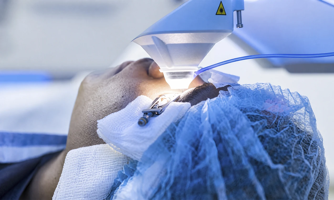
The procedure uses a femtosecond laser, the Zeiss VisuMax, to shape a small lenticule of tissue within the cornea, which is then removed through that small incision. Removing the lenticule changes the cornea’s refractive power and corrects the visual defect.
Why Is It Considered Minimally Invasive?
The key difference from LASIK is the incision. LASIK requires creating a corneal flap with an arc of about 20 millimeters to access the stroma, while SMILE works through an incision of just 2 to 4 mm, disrupting nearly 80% less corneal surface.
This preservation has several important clinical implications:
- Greater biomechanical integrity of the cornea, which matters for patients with high prescriptions or thin corneas.
- Lower incidence of post-operative dry eye, since fewer corneal nerves are affected during surgery.
- No risk of flap displacement from later trauma, a known (though uncommon) LASIK complication.
- Fast visual recovery: most patients notice significant improvement the day after surgery.
Clinical Indications
ReLEx SMILE is appropriate for patients with:
- Myopia of up to -10 diopters
- Myopic astigmatism of up to -5 diopters
- Age 18 or older, with a stable prescription for at least 12 months
- Adequate corneal thickness (confirmed by pre-operative pachymetry)
- Normal corneal topography, with no signs of keratoconus or ectasia
It is not currently recommended for pure hyperopia or presbyopia alone, though Zeiss has published research on these applications for future generations of the procedure.
Advantages Over Other Techniques
| Feature | ReLEx SMILE | Traditional LASIK | PRK |
|---|---|---|---|
| Incision size | 2-4 mm | ~20 mm (full flap) | Entire surface |
| Corneal flap | No | Yes | Not applicable |
| Functional visual recovery | 24 hours | 24-48 hours | 5-7 days |
| Post-op dry eye | Minimal | Common | Common |
| Return to contact sports | 1 week | 3-4 weeks | 3-4 weeks |
The Procedure, Step by Step
- Pre-operative evaluation: corneal topography, pachymetry, aberrometry, and visual quality analysis. Takes about 90 minutes.
- Day of surgery: topical anesthetic eye drops are applied. The patient lies down under the VisuMax femtosecond laser.
- Femtosecond laser: over 10 to 15 seconds per eye, the laser shapes the lenticule within the corneal stroma and creates the access incision.
- Lenticule extraction: the surgeon manually removes the lenticule through the small incision. This is the key clinical step.
- Natural closure: the incision closes on its own through natural tissue adhesion, with no stitches needed.
- Immediate observation: the patient rests for a few minutes and then leaves with post-operative instructions.
The entire procedure takes about 30 minutes for both eyes, though time under the laser itself is only 10 to 15 minutes per eye.
Recovery and Aftercare
For the first 4 to 6 hours, you may feel like there’s something in your eye or notice some tearing. That’s normal and resolves on its own. Antibiotic and anti-inflammatory eye drops are used during the first week, along with artificial tears as needed.
Significant visual improvement is noticeable the moment you wake up the next day. Vision continues to sharpen over the following 2 to 4 weeks until it fully stabilizes.
Recommendations for the first month:
- Avoid rubbing your eyes for at least 2 weeks
- No swimming or hot tubs for 2 weeks
- No intense exercise for the first 7 days
- Wear UV-protective sunglasses outdoors
Why Choose ReLEx SMILE at Centro Láser
Centro Láser was the first eye care center to introduce ReLEx SMILE in the Dominican Republic, with Zeiss VisuMax technology and a team of refractive surgeons trained internationally in the technique.
Our pre-operative protocol includes Pentacam corneal topography, ultrasonic pachymetry, and wavefront aberrometry, ensuring every patient meets the clinical criteria before surgery is scheduled.